Q: How have you used fresh allografts in new or innovative ways?
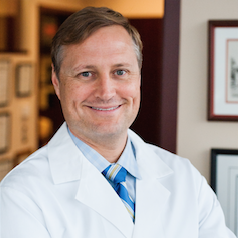
 Matthew Provencher, MD (Massachusetts General Hospital, Boston, Mass.): Traditionally, fresh osteochondral allografts have been used in and around the knee joint. They were first described for use in the condyles of the knee, but the indications have since expanded due to additional allograft availability, improved instrumentation, and demonstration of good long-term outcomes. I’ve expanded the allograft technology to use in multiple other joints, such as using ankle allograft tissue for shoulder reconstructive procedures. For example, we were the first to describe the use of a fresh distal tibial allograft for reconstruction of glenoid bone defects. This has been done since 2008 with very promising results both clinically as well as healing on computed tomography scan demonstrating solid incorporation.
Due to the favorable clinical outcomes of fresh distal tibial allograft to the glenoid, we have also started to utilize fresh talus allograft for large Hill-Sachs lesions as well as reverse Hill-Sachs injuries. The reason for utilizing talar allograft for the humerus is: 1) we have shown that the talus fits well for both typical Hill-Sachs and reverse Hill-Sachs lesions; 2) the ready availability of talus grafts (sometimes within one to two weeks); and 3) no need for size matching, much like the fresh distal tibia, which is also not size matched. The talus has to be cut in a special manner in order to accommodate the correct sizing and fit for the Hill-Sachs. We have found that the curvature, as well as potential for cartilage restoration particularly beneficial, especially in the setting of a reverse Hill Sachs.
After doing the sizing and contouring work, we found you have to make cuts in the correct area to obtain a congruent fit between talus and humerus. The curvature matches very well and since the talus is comprised of dense corticocancellous bone, this incorporates well and is a robust construct. We also utilize headless compression screws to ensure that the graft is compressed well after final fit is obtained.
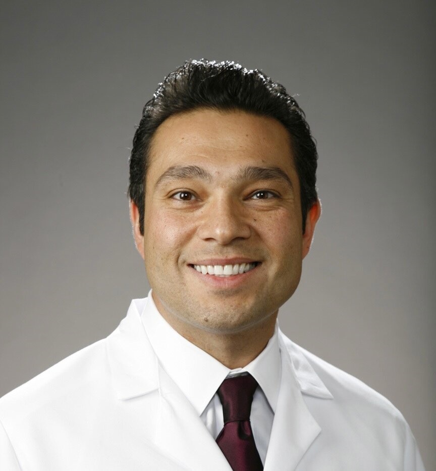
 Raffy Mirzayan, MD (Kaiser Permanente, Southern California): The trochlea and patella are two challenging areas to treat chondral injuries for surgeons. I typically see two types of patients with chondral loss in the patellofemoral joint. The first type are patients in their teens and early 20’s with recurrent patellar instability who have underlying trochlear dysplasia and have severe chondral loss of the patella. The second type are patients in their 40's who have isolated patellofemoral arthritis. In these cases, I resurface the entire patella by cutting off the damaged articular portion from the patient (similar to a total knee replacement) and obtaining a fresh patellar graft, cutting the articular surface off the donor, and then fixating it to the patient’s native patella with headless compression screws. The trochlea is typically transplanted with a 35 mm osteochondral graft from the donor who has been carefully screened to have a deep groove to address the trochlear dysplasia.
I also use fresh osteochondral allografts in the elbow for the treatment of osteochrondritis dissecans of the capitellum in overhead athletes, allowing them to return to throwing sports.
Scott Rodeo, MD (Hospital for Special Surgery, New York City, N.Y.): One of the significant advantages of these osteochondral allograft plugs is the "off-the-shelf" availability. On the rare occasion where patients with ACL injury have an actual osteochondral defect at the site of the translational contusion, the fresh allograft can be used to resurface this defect. Although this is uncommon, some patients will have such a deep defect.
Q: How do fresh osteochondral allografts affect patient care?
RM: Many patients come to me with little hope after having seen other physicians who gave them no options. These patients are in their 30s and 40s with arthritis and are told the only other option is an artificial knee replacement. This biologic solution frequently allows them to return to their regular activities and positively impacts their lives.
MP: It opens up more options for me. Fresh osteochondral allograft is FDA-approved for joints throughout the body and it has allowed more options and creative solutions for cartilage and/or bony impacts of the joint. The JRF Ortho products are available much sooner than others; instead of waiting three to six months for allografts, we wait two to three weeks at most.
Most defects are on the medial condyles. If everyone is requesting fresh medial condyle, availability becomes challenging. However, we conducted a study showing the lateral condyle, for most areas of the medial condyle, can work just as well. If I’m doing a knee I’ll ask for the first available condyle instead of waiting specifically for the medial. As long as the plug isn’t extremely huge — 25 mm or greater — we’ve found you can interchange the medial to lateral and lateral to medial and have the graft in a few weeks instead of a few months.
 SR: The availability of tissue allows treatment of such defects without the necessity to delay surgery to wait for availability of fresh osteochondral tissue.
Q: How can JRF Ortho encourage surgeons to use fresh allografts in new and innovative ways?
SR: The primary advantage will be the improved availability of tissue. This could allow us for indications where tissue transplant is less commonly used, such as for large Hill-Sachs lesion in the shoulder or capitellar defects in the elbow in athletes with osteochondritis dissecans.
MP: Think of areas where joints may be reasonably conserved. You are looking at cartilage bone solutions and you don’t have to use the same joint to reconstruct that joint and have a good outcome.
RM: JRF Ortho has been supportive of my innovative techniques using fresh grafts by making custom order grafts for me and ensuring they meet my patient’s needs. They have also credited me for the development of the BioPFJ™ technique. I am pleased to share this technique with other surgeons and improve patient outcomes.
This article is sponsored by JRF Ortho.
